
Description
GD/L69B Central Venous Catherization Simulator
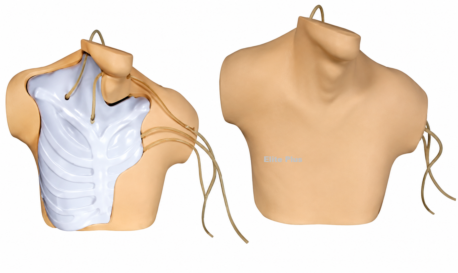
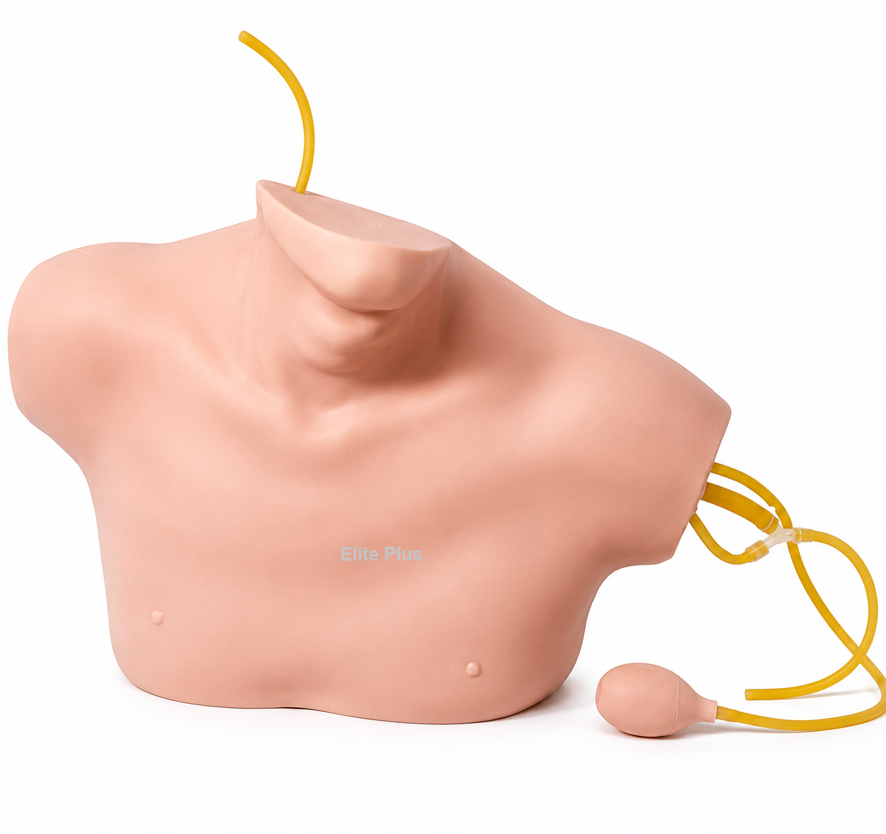
GD/L69B Central Venous Catherization Simulator is a realistic clinical skills training model for teaching central venous catheterization and venous-access procedures. Designed for medical colleges, nursing schools, hospitals, and simulation centres, this simulator helps learners practise vital techniques in a safe, controlled training environment.
The GD/L69B Central Venous Catherization Simulator presents clear cervical anatomical landmarks, including the sternal notch, clavicle, cervical ribs, and sternocleidomastoid muscle. These visible structures help learners identify relevant anatomical reference points during central venous access training.
This central venous catheterization trainer includes a detailed deep left-neck venous system with the common carotid artery, internal jugular vein, external jugular vein, subclavian vein, and brachiocephalic vein. It supports practice of internal jugular vein puncture and cannulation as well as subclavian vein puncture and cannulation.
The venous access training manikin can simulate a carotid artery pulse, providing a more realistic learning experience. Replaceable skin and veins support repeated classroom practice, while the simulated resistance and distinct “pop” sensation help learners recognise successful needle entry into the vein.
Explore Elite Plus nursing manikins, CPR training manikins, and ACLS manikins for further healthcare education and clinical simulation equipment.
Choose the GD/L69B Central Venous Catherization Simulator for dependable central line insertion practice, internal jugular cannulation training, and subclavian vein catheterization education. The GD/L69B Central Venous Catherization Simulator is an effective solution for modern clinical skills training.
Features:
1.Cervical anatomical landmarks are quite clear and indicate structures of the sternal notch , clavicle , right cervical ribs , sternocleidomastoid and etc.
2.Venous system in the deep part of left neck consists of common carotid artery, internal jugular vein, external jugular vein, subclinical vein and brachiocephalic vein.
3.Internal jugular vein puncture and cannulation can be exercised.
4.Subclinical vein puncture and cannulation can be exercised.
5.Carotid artery pulse can be simulated.
6.The skin and veins can be replaced and students will feel an apparent “pop” when the needle has been thrust into the vein.

Reviews
There are no reviews yet.